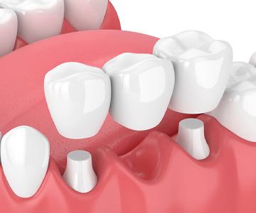
Treatment of edentulism (missing teeth) with a dental bridge is one of the most common and effective methods. A dental bridge allows for the restoration of both chewing function and smile aesthetics. It is a fixed prosthetic construction that is supported by healthy adjacent teeth and fills the space of a missing tooth. In this article, we will discuss what a dental bridge is, the types available, and how the placement process is carried out.
To book a visit, sign up for a consultation. To clarify the details, our operator will contact you.

3D computed tomography in dentistry
07 November 2024
Innovative technologies help us deliver high-quality, accurate, and fast services to our patients, which gives our clinic a competitive edge in the market. When discussing innovative approaches, it’s essential to highlight the significance of 3D computed tomography. Check out our blog to learn what 3D CT is and why it’s necessary for creating an effective treatment plan.
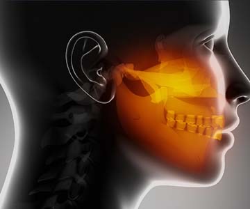
3D computed tomography (3D CT or X-ray) is used in nearly all dental services to plan treatments and gather complete information about the patient’s oral cavity. It provides dentists with a detailed and comprehensive view of the patient’s upper and lower jaw, right and left joints, and sinus cavity.
Additionally, 3D computed tomography enables us to:
- Identify hidden sources of pain: Often, the cause of tooth pain is unknown or cannot be detected with a 2D X-ray. However, with modern 3D CT, even the smallest damage is clearly visible.
- Plan procedures accurately, such as tooth extractions, as the 3D X-ray shows the shape of the tooth root and nerve positioning.
- Evaluate bone quality for implants, including its height and width, to determine if there is enough bone for implant placement.
- Examine the unique anatomy and inflammation in root canals before root canal treatment.
- Assess any damage in the temporomandibular joint. If a patient feels pain while chewing or at rest around the ear area, these symptoms may indicate a chewing system disorder.
- Monitor the teeth of adolescents, assess their bite, and plan suitable treatment using both 2D and 3D images.
Blits Dental - Kakhaber Kharebava clinic uses the highest-quality 3D CT device, the “Orthophos SL,” manufactured by the German company Dentsply Sirona. With the Orthophos SL 3D device, dental pathologies are diagnosed through all-in-one virtual visualization, allowing for comprehensive software modeling. In a single scan, we can capture the full upper and lower jaw, right and left joints, and highmore hollow. The information provided by the Orthophos SL 3D device is valuable for both dental and ENT (ear, nose, and throat) treatments.
As we approach the end of the blog, you might be curious about how the procedure is conducted. Before starting the CT scan, it’s important to remove any metal objects from your face and neck, such as necklaces, earrings, glasses, or other accessories. The X-ray is done in a standing position, and you should remain as still as possible to avoid a blurry image. During the scan, the scanner rotates 360 degrees around your head for about a minute, capturing hundreds of 2D images that are then converted into a 3D model using computer software.
Our clinic is always dedicated to ensuring your comfort
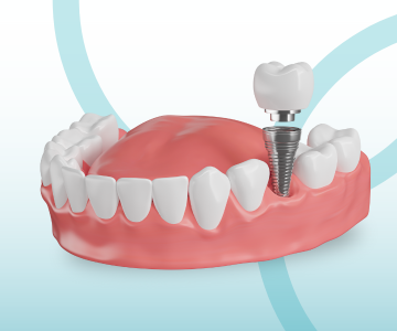
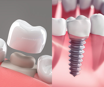
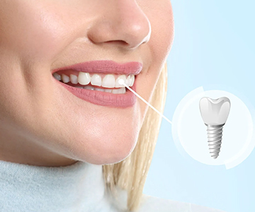
Missing teeth are no longer a problem! Dental implants offer the most advanced, durable, and natural-looking solution for restoring your smile. An implant, together with its crown, mimics a real tooth both aesthetically and functionally-helping you regain confidence, comfort, and a complete smile.
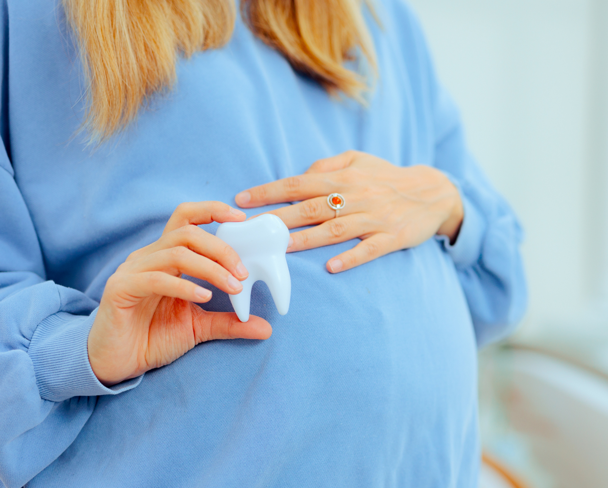
During pregnancy, hormonal changes can cause gum inflammation, bleeding, enamel erosion, and an increased risk of cavities. That’s why visiting the dentist during pregnancy is especially important.
Gnathology is one of the leading branches of 21st-century dentistry. It forms the foundation for any complex dental treatment planning
Tooth loss (edentulism) affects not only the appearance of your smile but also the overall functional health of your oral cavity
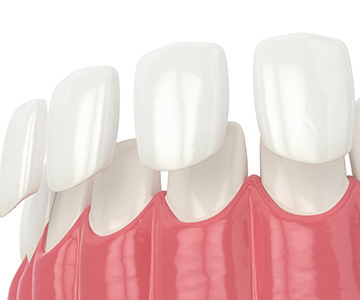
Dental veneers can be made from various materials, but ceramic (porcelain) veneers are the most widely used.
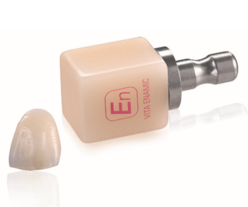
Modern aesthetic and functional dentistry is continually evolving, striving to identify restorative materials that combine exceptional strength
The eruption of baby teeth is one of the most important stages in a child’s early development.
Modern dentistry increasingly emphasizes the importance of orthodontic care.
Oral health care begins long before the first permanent tooth erupts.
A smile is one of the key elements of a person’s visual identity. It conveys confidence and positivity. However, the beauty of a smile is not only an aesthetic factor—it is directly connected to oral health.
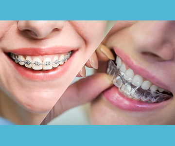
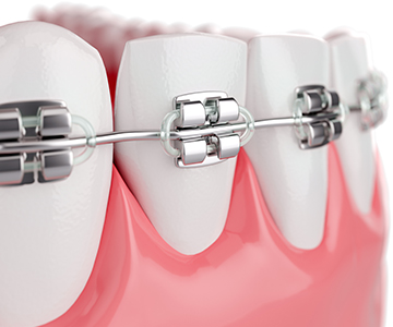
Orthodontic treatment has long gone beyond the limits of traditional metal braces.
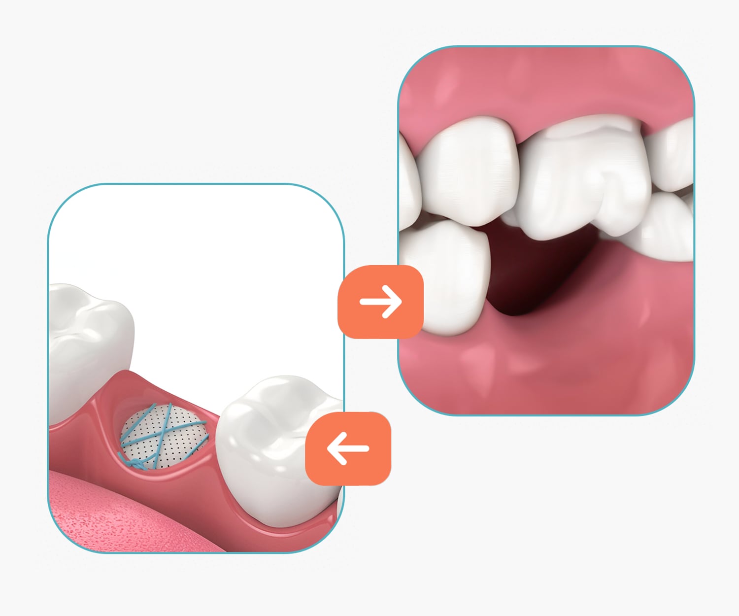
Dental implantation is the best method for restoring missing teeth. However, for the procedure to be successful, the jawbone must have sufficient volume and density.
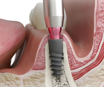
Dental implantation is one of the most effective and safest surgical procedures in modern dentistry for restoring missing teeth.
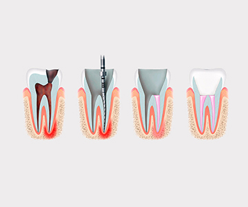
Initial endodontic (root canal) treatment is usually successful and helps preserve the natural tooth.
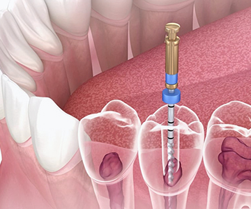
Root canal treatment, also known as endodontic therapy, is one of the most frequently discussed yet often misunderstood dental procedures.
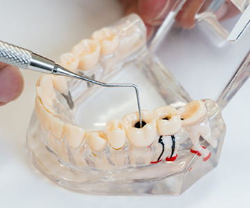
Tooth decay is one of the most common dental conditions, involving damage to the hard tissues of the teeth
Modern dentistry is constantly evolving, offering improved methods for solving complex issues.
Today, there are numerous teeth whitening options—both at home and professionally done.


.jpeg)
.jpeg)